Follow us
Latest News & Events


How to choose a digital pathology slide scanner?
Digital pathology has transformed how pathologists view, analyze, and manage pathology slides. Instead of relying only on traditional microscopes, laboratories can now digitize entire glass slides for faster diagnosis, remote collaboration, and AI-assisted analysis.
A digital pathology slide scanner is the core device that makes this transformation possible.
What is a digital pathology slide scanner?
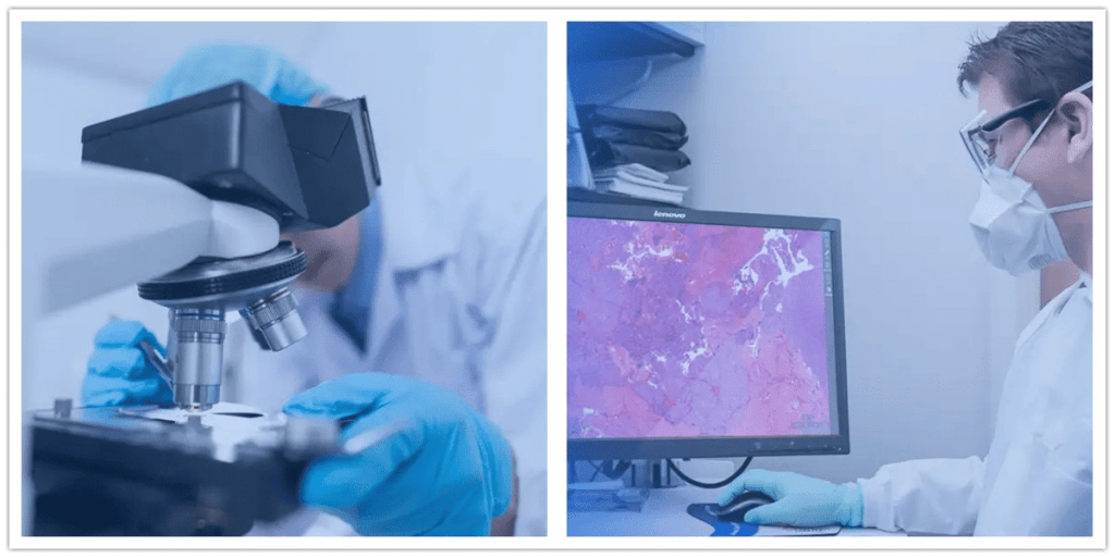
A digital pathology slide scanner is an imaging system used in pathology, clinical medicine, biological research, and life sciences. It automatically converts glass slides into high-resolution digital images for viewing, analysis, and long-term storage.
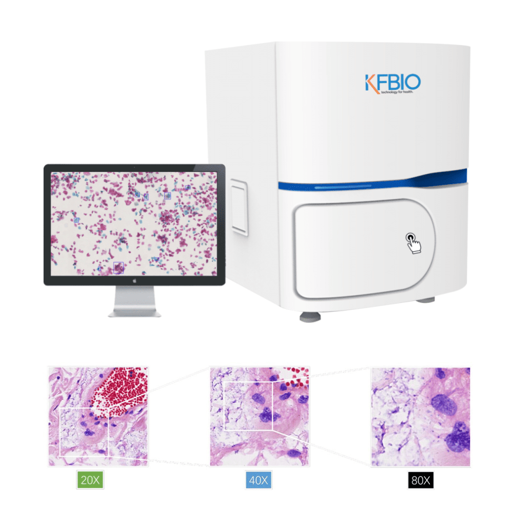
Compared with traditional microscopes, a digital pathology scanner captures whole-slide images automatically. It uses high-precision image stitching to produce seamless digital slides. In addition, K-SCP color processing technology accurately restores the original tissue colors.
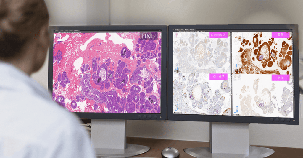
The scanner stores and compresses image data into electronic files. Medical professionals, researchers, and engineers can easily view, zoom, measure, and annotate these digital slides with dedicated viewing software.
As a result, digital pathology scanners provide a strong technical foundation for remote pathology consultation, telepathology, education, and AI-assisted pathology diagnosis.

Why Choose a Digital Pathology Slide Scanner Instead of a Traditional Microscope?
Digital pathology scanners offer significant advantages over conventional microscopes. They improve workflow efficiency, image consistency, and diagnostic accuracy.
Below are the key benefits.
1. Faster Scanning Speed
Traditional microscopes require manual operation. As a result, slide review speed depends heavily on the pathologist’s experience and workload. In addition, only one slide can be examined at a time.
By contrast, a digital pathology slide scanner performs high-throughput scanning automatically. KFBIO digital pathology scanners can scan a slide in as little as 25 seconds and process up to 400 slides in a single batch.
Therefore, laboratories can significantly improve efficiency while reducing manual workload.
2. More Stable and Quieter Operation
Traditional microscope performance varies for several reasons. Different microscopes, operating environments, and users may produce different scanning results. Consequently, image consistency can become a challenge.
Digital pathology scanners automate the scanning process and generate high-resolution whole-slide images with consistent quality.
Moreover, KFBIO digital pathology scanners use magnetic motor control technology. This design minimizes mechanical wear and eliminates backlash during operation. As a result, scanning becomes smoother, quieter, and more reliable.
The slide scanning success rate reaches 99.85%, helping laboratories achieve stable and repeatable digital imaging.
3. Automatic Scanning with Intelligent Analysis
Traditional microscopes require continuous manual operation. Pathologists must review every slide individually and rely on their expertise to complete the analysis. Therefore, results may be influenced by subjective interpretation.
Digital pathology scanners simplify this workflow with one-click automated scanning. They can process large numbers of slides with minimal manual intervention, allowing pathologists to focus on diagnosis instead of repetitive scanning tasks.
Furthermore, KFBIO AI solutions support intelligent image analysis through deep learning models trained on large, annotated pathology slide databases. These AI tools assist pathologists by improving efficiency and supporting more consistent diagnostic workflows.
4. High Resolution and Excellent Color Fidelity
Traditional microscopes provide excellent optical resolution. However, viewing an entire slide at high magnification can be difficult and time-consuming.
Digital pathology scanners overcome this limitation by creating complete whole-slide images. Their highest resolution can reach approximately 0.125 μm/pixel.
The scanner uses high-precision seamless image stitching to reconstruct the original specimen with exceptional accuracy. It compares image gradient ratios against preset values to verify stitching quality. This process ensures objective image evaluation and optimal stitching performance.
In addition, KFBIO digital pathology scanners use proprietary K-SCP color processing technology. This technology enhances color saturation, color temperature, and image contrast while maintaining accurate tissue appearance.
The system also compensates for color differences across monitors. As a result, pathologists can view highly faithful digital images regardless of the display device.
Overall, high-resolution imaging and accurate color reproduction help improve diagnostic confidence and support reliable digital pathology workflows.
Key Factors to Consider When Choosing a Digital Pathology Scanner
Choosing the right digital pathology scanner is essential for building an efficient digital pathology workflow. Different scanners offer different performance levels, scanning technologies, and imaging capabilities.
Before making a decision, evaluate the following key factors.
1. Scanning speed
Scanning speed directly affects laboratory efficiency. A faster scanner can process more slides each day and shorten turnaround times.
You can also estimate daily scanning capacity based on the scanner’s speed. This helps laboratories plan workloads more effectively and improve operational efficiency.
However, scanning speed should not be the only consideration. Image quality and scanning stability are equally important.
2. Scanning Mode
The scanning mode determines image quality, scanning stability, operating noise, and overall workflow efficiency.
Modern digital pathology scanners generally support one or more of the following scanning modes:
- Flat whole-slide scanning
- Z-stack scanning
- Extended focus scanning
Each mode is designed for different applications.
(1) Flat whole-slide scanning:
Flat whole-slide scanning is the most common scanning method.
The scanner captures a large number of square image tiles across the entire slide. During scanning, adjacent tiles typically overlap by 2%–5%. The software then stitches these images together to create one seamless whole-slide image.
This method delivers fast scanning and excellent image quality for standard pathology slides.
However, it works best with flat, single-layer specimens. Thick or uneven slides may produce out-of-focus areas that reduce image quality.
(2) Z-Stack scanning:
Z-stack scanning captures images at multiple focal planes along the vertical (Z) axis.
A precision servo motor moves smoothly through different focus positions without vibration. The scanner captures multiple image layers and combines them into high-quality multi-planar images.
As a result, pathologists can observe tissue structures at different depths, similar to adjusting the fine focus on a conventional microscope.
Compared with flat scanning, Z-stack scanning offers several advantages:
- Fewer image tiles are required, simplifying image stitching.
- Multiple focal layers capture more tissue information.
- Deconvolution algorithms generate clearer single-layer images.
- Uneven or thick tissue sections can be scanned more accurately.
Therefore, Z-stack scanning is particularly valuable for cytology, hematology, and other applications requiring multiple focal planes.
(3)Extended focus scanning:
Extended focus scanning combines several images captured at different focal planes into a single sharp composite image.
Compared with full multi-layer virtual images, this method offers two major advantages:
- Faster image acquisition
- Smaller file sizes
As a result, extended focus scanning provides a practical balance between image clarity, scanning speed, and storage requirements.
Mainstream product comparison
| Brand | KFBIO | LEICA | 3DHISTECH | MOTIC |
|---|---|---|---|---|
| Scan mode | Multilayer scanning (Z-stack) | Multilayer scanning (Z-stack) | Multilayer (Z-stack) and extended focus scanning | Multilayer scanning (Z-stack) |
Overall, laboratories should choose a scanning mode based on their diagnostic requirements, slide characteristics, and daily workload. While flat scanning is sufficient for many routine applications, Z-stack and extended focus scanning provide greater flexibility for more complex specimens.
3.Objective lens
The objective lens is one of the most important components of a digital pathology scanner. Its optical performance directly affects image quality, scanning accuracy, and diagnostic reliability.
High-quality objective lenses produce sharper images, better color reproduction, and greater detail. As a result, they play a critical role in digital pathology.
Leading objective lens manufacturers include Olympus, Zeiss, Nikon, and Leica.
KFBIO digital pathology scanners use premium Olympus dual objective lenses together with an advanced optical filter system. The narrow-band single-pass filter ensures consistent spectral transmission while minimizing cross-color interference.
The filter system provides:
- Up to 99% light transmittance
- OD6 cut-off frequency
- High image contrast
- High brightness
- Zero color drift
- Accurate color reproduction
Together, these technologies deliver stable and high-quality whole-slide images.

Key Objective Lens Specifications
When evaluating an objective lens, focus on the following three specifications:
- Resolution
- Numerical Aperture (NA)
- Magnification
Each specification influences image quality in a different way.
Resolution
Resolution describes the minimum distance between two adjacent points that can still be distinguished as separate objects.
Simply put, the smaller the minimum distance, the higher the resolution.
For comparison:
- The human eye resolves approximately 100 μm.
- A conventional optical microscope resolves about 0.2 μm.
- KFBIO digital pathology scanners achieve a minimum resolution of 0.25 μm.
Higher resolution allows pathologists to observe finer cellular structures and tissue details.
Numerical Aperture (NA)
Numerical Aperture (NA) measures an objective lens’s ability to gather light and resolve fine details.
The relationship between NA and resolution is shown below:
δ = 0.61λ / NA
Where:
- δ = minimum resolvable distance
- λ = wavelength of light (approximately 0.555 μm for white light)
- NA = numerical aperture
A larger NA produces higher resolution and reveals more microscopic details.
In general:
- Higher NA → Higher resolution
- Higher NA → Better image quality
- Higher NA → Greater diagnostic confidence
Different objectives have different NA values.
Typically:
- Dry objectives: NA 0.05–0.95
- Oil immersion objectives: NA 0.85–1.40
An NA of 0.95 is considered excellent for a dry objective.
KFBIO objective lenses achieve this high-performance level.
Magnification
Magnification refers to the ratio between the final image size and the actual specimen size.
For traditional microscopes, total magnification equals:
Objective magnification × Eyepiece magnification
However, magnification alone does not determine image quality. Resolution ultimately limits the amount of visible detail.
For digital pathology, extremely high magnification is rarely necessary. Most pathology applications focus on observing cells and tissue morphology rather than extremely small structures.
Today, the most common magnifications are:
- 20×
- 40×
- 80×
These magnifications provide an excellent balance between image detail and scanning efficiency.
Mainstream product comparison
| Brand | KFBIO | LEICA | MOTIC |
|---|---|---|---|
| Objective lens | OLYMPUS apochromat objective lens | Aperio GT,Custom-made by Leica Microsystems | Plan APOCHROMAT |
| Numerical Aperture | 20X, NA 0.8 40X, NA 0.95 | 20x, NA 0.75 | 20X,NA 0.75 |
| Resolution | ≤0.5 µm/pixel(20X) ≤0.25 µm/pixel(40X) | 0.26 µm/pixel(40X) | 0.52µm/pixel(20X) 0.26µm/pixel(40X) |
| Magnification | 20X/40X/80Xx | 1.25X/5X/10X/20X/40X/63X | 20X/40X |
Illumination
Illumination determines which applications a digital pathology scanner can support.
Most modern scanners provide one or both of the following imaging modes:
- Bright-field imaging
- Fluorescence imaging
Bright-Field Imaging
Bright-field imaging is the standard mode used in routine pathology.
Light passes directly through the specimen and enters the objective lens. As a result, the background appears bright while stained tissue structures become clearly visible.
This mode is suitable for most H&E and routine histopathology applications.
Fluorescence Imaging
Fluorescence imaging detects fluorescent signals emitted by labeled specimens.
It allows pathologists and researchers to observe the distribution of specific cells, DNA sequences, RNA molecules, or biomarkers after fluorescent staining.
Therefore, fluorescence imaging is widely used in molecular pathology, cytogenetics, and biomedical research.
Mainstream product comparison
| Brand | KFBIO | LEICA | 3DHISTECH |
|---|---|---|---|
| Illumination | Bright-field & Fluorescence | Bright-field & Fluorescence | Bright-field & Fluorescence |
Supporting both imaging modes gives laboratories greater flexibility across clinical diagnosis and scientific research.
KFBIO digital pathology scanners support seamless switching between bright-field and fluorescence imaging. In addition, the independent optical path design improves scanning stability and overall efficiency.
The bright-field system uses a professional 3CCD linear camera, while the fluorescence system uses a scientific-grade sCMOS camera.
Independent RGB processing channels provide:
- More accurate color reproduction
- Higher image clarity
- Better tissue visualization

Capacity
Capacity refers to the number of slides that a scanner can process in one batch.
Generally, a larger capacity improves laboratory efficiency. However, high-capacity scanners also require a greater investment.
Therefore, laboratories should choose a scanner that matches their daily slide volume and workflow requirements.
Mainstream product comparison
| Brand | KFBIO | LEICA | 3DHISTECH |
|---|---|---|---|
| Capacity | 2/5/20/40/120/400 | 120/400/450 | 1/11/12/150/250/1000 |
KFBIO offers digital pathology scanners for a wide range of applications.
These include:
- Low-throughput scanners for remote pathology
- High-throughput scanners for fully digital pathology laboratories
- Bright-field scanners for routine diagnosis
- Bright-field and fluorescence scanners for advanced research
As a result, laboratories of different sizes can choose a solution that best fits their clinical and research needs.

Conclusion
Selecting the right digital pathology scanner requires more than comparing specifications.
Laboratories should evaluate scanning speed, scanning mode, objective lens performance, illumination options, and scanner capacity together. These factors directly affect workflow efficiency, image quality, and long-term scalability.
KFBIO provides a complete portfolio of digital pathology scanners designed for hospitals, pathology laboratories, and research institutions. Whether your goal is routine diagnosis, remote consultation, AI-assisted pathology, or scientific research, KFBIO offers flexible solutions to support every stage of digital transformation.